3D ultrasound imaging is one of the most desired features of an ultrasound imaging system. It provides comprehensive evaluations of the targeted tissue and effectively alleviates the user...
3D ultrasound imaging is one of the most desired features of an ultrasound imaging system. It provides comprehensive evaluations of the targeted tissue and effectively alleviates the user dependence of ultrasound. Achieving a high imaging volume rate is critical for all the 3D ultrasound applications. The current 3D ultrasound technologies use 1D or 2D ultrasound transducers, but they face challenges including limited imaging volume rate, high cost, and technical issues.
A high volume-rate 3D ultrasound imaging method and a device based on Fast Acoustic Steering via Tilting Electromechanical Reflectors (FASTER). This technology addresses challenges of conventional 3D ultrasound imaging like high cost, suboptimal imaging quality, and low volume scan rate. FASTER is capable of high volume rate (up to 500 Hz compared to 0.2-20 Hz for conventional techniques) large field-of-view 3D imaging with conventional 1D transducers.
The super-resolution ultrasound microvessel imaging (SR-UMI) technique offers a promising solution to the challenge of achieving both high imaging resolution and deep...
The super-resolution ultrasound microvessel imaging (SR-UMI) technique offers a promising solution to the challenge of achieving both high imaging resolution and deep penetration in biomedical imaging. By using ultrasound contrast microbubbles and super-resolution strategies similar to PALM and STORM, SR-UMI enhances ultrasound imaging resolution by tenfold while maintaining deep penetration. This technique can resolve capillary-scale blood vessels at depths greater than 10 cm and measure microvascular blood flow speeds as low as 1 mm/s. SR-UMI provides detailed structural and functional information about tissue microvasculature and is noninvasive, low-cost, and free of ionizing radiation. Despite its potential, SR-UMI faces challenges such as slow data acquisition and computationally intensive post-processing, which hinder its clinical translation.
This technology is a method of real-time Super-Resolution Ultrasound Microvessel Imaging (SR-UMI). Current SR-UMI requires hours of data post-processing, making it impractical for clinical, diagnostic applications. Implementing advances in deep learning and parallel computing enabled real-time microbubble signal extraction, separation, localization, tracking, and quantitative analysis and display. This technology has a wide range of clinical applications, including but not limited to, the diagnosis and characterization of a range of disorders (e.g., cancer, cardiovascular disease, and neurological diseases).
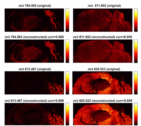
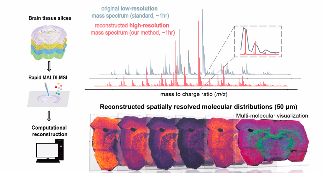
Researchers have developed a method that accelerates data acquisition for Fourier transform-ion cyclotron resonance (FT-ICR) mass spectrometry imaging (MSI) by ten folds...
Researchers have developed a method that accelerates data acquisition for Fourier transform-ion cyclotron resonance (FT-ICR) mass spectrometry imaging (MSI) by ten folds while maintaining high mass and spatial resolution and accuracy. Compared to other methods in MSI data acquisition/reconstruction, this approach exploits redundancy in the data, eventually reducing the time for data collection. The primary applications of the technology will be in clinical diagnostics, drug metabolism studies and localization/characterization of biomolecules within tissue samples.
Radiological clips, or markers, are inserted at the time of biopsy to mark tumor locations, lesions and lymph nodes for consistent identification over time and multiple...
Radiological clips, or markers, are inserted at the time of biopsy to mark tumor locations, lesions and lymph nodes for consistent identification over time and multiple treatments. Prof. Michael Oelze has developed a new type of radiological clip that have a unique ultrasonic signal, acting like both a beacon and a barcode. This technology can be particularly helpful to mark multiple areas that are close together.