

Prof. Mary Kraft and her research group have developed a computational reconstruction strategy to reshape 3D images acquired from depth profiling mode on secondary ion mass spectrometry (SIMS). This strategy can enhance understanding of structure function relationship of materials using SIMs (e.g. subcellular biological processes). For samples with nonplanar surfaces, secondary ions detected in the same SIMS depth profiling image, and thus depicted at the same z-position with respect to the surface may be from molecules with different z positions. 3D SIMS image depth correction strategy is needed when both substrate signals and atomic force microscopy data are not available for NanoSIMS depth profiling. The reconstruction strategy accurately captures the basic shape of the cell as well as the surface features, in addition to reducing time for complementary instrumental data collection.
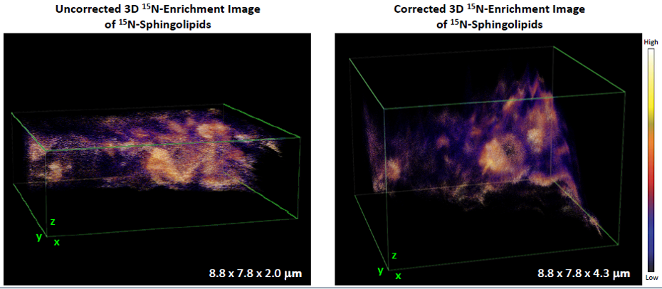
Figure 1. Comparison of 3D 18O-Enrichment 3D SIMS Images of 18O-Cholesterol (left: uncorrected, right: corrected with the reconstruction strategy)