

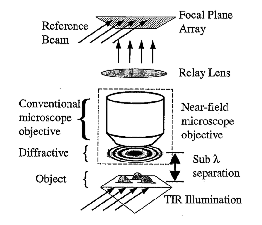
A near-field microscope using one or more diffractive elements placed in the near-field of an object to be imaged. A diffractive covers the entire object, thus signal may thereby be gathered from the entire object, and advantageously increase the signal-to-noise ratio of the resulting image, as well as greatly improve the acquisition speed. Near-field microscopy overcomes the limitation of conventional microscopy in that subwavelength and nanometer-scale features can be imaged and measured without contact.
Conventional Near-field Scanning Optical Microscopes (NSOM) and Atomic Force Microscopes (AFM) used for collecting images of sub-wavelength features in nanostructures and biological samples are inherently slow and can damage the object they are imaging. Their resolution is limited by the aperture of the scanning tip; hence a low signal-to-noise ratio is achieved. This Near-field Diffractive Microscope (NDM) addresses these issues by collecting data from the entire scattering surface simultaneously without the need for a small aperture to achieve high resolution images. The Near-field Diffractive Microscope uses a Fresnel plate as a single diffractive element to provide all the spatial frequencies needed to collect exhaustive information from different regions of the sample.
This optical device provides several benefits over the existing instruments, including increased speed of data collection and enhanced resolution. It has the potential to be of use in the imaging of samples from various fields - from life sciences to material science and in microlithography - where the ability to resolve atomic scale features with great speed and accuracy are crucial.